(途中)ヒトサイトメガロウイルス誘発特異的1番染色体破壊

Fortunato, E. A., Dell’Aquila, M. L., & Spector, D. H. (2000). Specific chromosome 1 breaks induced by human cytomegalovirus. Proceedings of the National Academy of Sciences of the United States of America, 97(2), 853. https://doi.org/10.1073/PNAS.97.2.853
要約
ヒトサイトメガロウイルス(HCMV)はウイルス誘発性先天性欠損症の主たる原因であり、免疫不全患者に深刻な問題を引き起こす。ここに、細胞周期のS期にHCMVに感染した細胞は、一番染色体の二つの部位:1q42と1q21が特異的に破壊されることを示す。また、感染細胞上清だけでなく、純粋化ビリオンが損傷の原因だと証明する。この特異的な破壊は線維芽細胞やHCMVの株を変えても生じる。ウイルスを中和抗体で培養すると染色体破壊の誘発を防げる。しかし、紫外線で不活化したウイルスは、未処理ウイルスと同程度の効率で1番染色体を特異的に破壊する。従って、ウイルスの吸着/浸透が不可欠だが、新たなウイルス遺伝子の発現は必要ない。このHCMVを介した、活発な分裂中の細胞における部位特異的損傷の誘導は、先天性感染児における神経学的欠損の発生の手掛りとなる可能性がある。
序章
多くのヒトウイルス(アデノウイルス/単純ヘルペス1・2型ウイルス/帯状疱疹ウイルス/エプスタインバーウイルス/ヒトサイトメガロウイルス(HCMV)/B型肝炎ウイルス/流行性耳下腺炎ウイルス/麻疹ウイルス/風疹ウイルス/ポリオウイルス/パピローマウイルス)[1-4]が非特異的な染色体異常や有糸分裂装置の破壊の原因となる多くの証拠がある。染色分体の切断および染色体粉砕が最も頻繁に観察される異常だが、転座 、コイリング欠損 、過剰凝縮 なども報告されている。損傷の大半は一見ランダムであり、発癌性アデノウイルスを除き、部位特異的な切断の証拠はない。アデノウイルス12型の場合、ヒト細胞への低倍数感染は、4つの異なる遺伝子座を特異的に脆弱にする[5,6]。最も頻繁な3つの部位:17q21-22、1p36、1q12-22は主要な核内低分子RNA遺伝子座と一致し、4つ目の部位である1q42-43は感染したヒト初代培養細胞では低頻度にしか観察されないが、5S rRNA遺伝子座に隣接している[5-10]。
[1]Harnden, D. G. (1974) in Chromosomes and Cancer, ed. German, J. (Wiley, New York), pp. 151–190.
[2]G. Lüleci, M. Sakízlí, and A. Günalp, “Selective chromosomal damage caused by human cytomegalovirus,” Acta Virol, vol. 24, no. 5, pp. 341–345, Sep. 1980, [Online]. Available: https://www.digitalniknihovna.cz/cvti/view/uuid:4c1b1eea-4705-441b-8a09-5f333b9d8031?page=uuid:2b29f6d6-a198-4890-a7a2-913be41206c5
[3]S. Abubakar, W. W. Au, M. S. Legator, and T. Albrecht, “Induction of chromosome aberrations and mitotic arrest by cytomegalovirus in human cells,” Environmental Mutagenesis, vol. 12, no. 4, pp. 409–420, 1988, doi: 10.1002/em.2860120409.
[4]C. Z. Deng et al., “Cytomegalovirus-enhanced induction of chromosome aberrations in human peripheral blood lymphocytes treated with potent genotoxic agents,” Environmental and Molecular Mutagenesis, vol. 19, no. 4, pp. 304–310, 1992, doi: 10.1002/em.2850190407.
[5]H. Zur Hausen, “Induction of specific chromosomal aberrations by adenovirus type 12 in human embryonic kidney cells,” J Virol, vol. 1, no. 6, pp. 1174–1185, Dec. 1967, doi: 10.1128/JVI.1.6.1174-1185.1967.
[6]D. M. Steffensen, P. Szabo, and J. K. McDougall, “Adenovirus 12 uncoiler regions of human chromosome 1 in relation to the 5S rRNA genes,” Exp Cell Res, vol. 100, no. 2, pp. 436–439, Jul. 1976, doi: 10.1016/0014-4827(76)90176-2.
[7]V. Lindgren, L. B. Bernstein, A. M. Weiner, and U. Francke, “Human U1 small nuclear RNA pseudogenes do not map to the site of the U1 genes in 1p36 but are clustered in 1q12-q22,” Mol Cell Biol, vol. 5, no. 9, pp. 2172–2180, Sep. 1985, doi: 10.1128/mcb.5.9.2172-2180.1985.
[8]V. Lindgren, M. Ares, A. M. Weiner, and U. Francke, “Human genes for U2 small nuclear RNA map to a major adenovirus 12 modification site on chromosome 17,” Nature, vol. 314, no. 6006, pp. 115–116, Mar. 1985, doi: 10.1038/314115a0.
[9]S. Schramayr, D. Caporossi, I. Mak, T. Jelinek, and S. Bacchetti, “Chromosomal damage induced by human adenovirus type 12 requires expression of the E1B 55-kilodalton viral protein,” J Virol, vol. 64, no. 5, pp. 2090–2095, May 1990, doi: 10.1128/jvi.64.5.2090-2095.1990.
[10]P. D. Sørensen, B. Lomholt, S. Frederiksen, and N. Tommerup, “Fine mapping of human 5S rRNA genes to chromosome 1q42.11----q42.13,” Cytogenet Cell Genet, vol. 57, no. 1, pp. 26–29, 1991, doi: 10.1159/000133107.
以上の観察結果は、ヒト疾患のウイルス媒介突然変異誘発の役割に関する重要な問題を提起している。本稿では、 先天性欠損症の主要なウイルス性の原因であるヘルペスウイルスことHCMVに焦点を当てる。毎年、新生児の約1%が先天性感染しており、その内5~10%は難聴、精神遅滞、失明、小頭症、脳石灰化[11-14]などの深刻な神経障害の兆候を出生時点で示している。更に、感染乳児の10~15%は出生時点で無症候性だが、その後様々なレベルの感音性難聴や学習障害を発症する。HCMV感染は、免疫不全患者にとっても大きな医学的問題である[11]。
ここ数年で、HCMVが宿主細胞の機能を著しく下方修正し、細胞周期の移行を阻害することが明らかになってきた[15-18]。先ず、HCMVが細胞内へ侵入すると、ホルモンや成長因子による修正の際に起こるのと同様のセカンドメッセンジャー型の反応を引き起こす[19]。生産的なHCMV感染は細胞のDNA複製の準備に関与するタンパク質群をコードする遺伝子発現を刺激し、許容細胞(※)で完全に「活性」状態になる[20-24]。HCMVはp53の定常状態のレベルを上昇させるが[15, 25, 26]、その下流の損傷応答標的であるp21やMDM2へのシグナル伝達はないようである[refs. 17 and 27, and E.A.F., 未発表データ]。このシグナル伝達のブロックが、ウイルスタンパク質の結合や阻害[25, 28, 29]やp53の隔離[30]であれ、HCMVが感染細胞を完全に「活性化」し、細胞死を誘発することなく遺伝毒性作用を起こすメカニズムを示唆している。
許容細胞(permissive cell)
ウイルスが感染し,増殖することができる細胞。ウイルスが許容細胞に感染すると,初期遺伝子群が発現し,細胞は細胞周期のS期に誘導され,ウイルスゲノムの複製が開始する。後期遺伝子群の発現によりウイルスの構造蛋白質が作られ,ウイルス粒子が細胞外に放出される。通常,許容細胞はウイルスの自然宿主に由来する。ウイルスが感染しても増殖できない細胞を非許容細胞という。非許容細胞はウイルス感染で形質転換を起こすことがある
HCMVに感染した培養液では、ランダムに分布する染色分体切断やギャップの数が有意に増加することが、これまでに何人かの研究者によって指摘されているが、特異的な切断は報告されていない[2-4]。しかし、これらの初期の研究では実験デザインに問題があり、特異的な染色体異常が観察されなかった可能性がある。この種のDNA損傷を検出するには、有糸分裂期の染色体を調べる必要がある。初期の実験の一部は、細胞をG0期に同期させた後に感染させている。前述の通り、これらの条件では細胞のDNA合成と分裂が阻害され、解析に必要な分裂中期の細胞は僅かしか得られない[15-18]。更に、これまでの実験の大半は低い感染倍率(moi, 1)で実施され、ウイルスの不均一な分布により初期の感染ラウンドが複雑化していた。最後、解析用の細胞が、ウイルスのDNA合成がピークに達して細胞分裂ができず、有史分裂像を示すことのできない感染後(pi)(48時間以降)の遅い時期に採取していた[2-4]。
G0からの放出時の感染とは異なり、S期に感染したヒト線維芽細胞はウイルスタンパク質の発現に抵抗性である[27]。The majority of the S-phase-infected cells move through mitosis and by 24 h pi (hpi) are back in G1, where viral gene expression can then initiate. If DNA damage occurred in this cycling population, the cells could divide before virus replication, resulting in one of the daughter cells being free of viral genomes. This daughter cell could be a reservoir for genetic damage, opening up the possibility that disease syndromes might stem from this early damage rather than be because of active viral replication and cell lysis in the infected individual. As the target cells in the developing fetus are likely still dividing, we have revisited the question of HCMV-induced damage by using S-phase infected cells as a model system.(27)。 S期に感染した細胞の大部分は有糸分裂を経て、24時間π期(hpi)にはG1に戻り、そこでウイルス遺伝子の発現が始まる。 この循環集団でDNA損傷が起こった場合、細胞はウイルス複製前に分裂し、その結果、娘細胞の一つはウイルスゲノムを持たない。 この娘細胞は遺伝的損傷のリザーバーとなる可能性があり、疾患症候群が感染者の活発なウイルス複製や細胞溶解のためではなく、この初期の損傷に起因する可能性が出てくる。 発育中の胎児の標的細胞はまだ分裂を続けている可能性が高いので、我々はS期感染細胞をモデル系として、HCMVによる損傷の問題を再検討した。
実験材料と方法
-細胞とウイルス
Primary human foreskin fibroblasts (FFs) were obtained from the University of California, San Diego Medical Center, and human embryonic lung cells were obtained from the American Type Culture Collection (ATCC) (no. CCL 137). Both cell types were propagated in MEM Earle media in incubators maintained at 37°C and 5% CO2. Media was supplemented with 10% heat inactivated FBS, L-glutamine (2 mM), penicillin (200 unitsyml), streptomycin (200 mgyml), amphotericin B (1.5 mgyml), and gentamycin sulfate (50 mgyml). The Towne (no. VR 977) and AD169 (no. VR 538) strains of HCMV were obtained from the ATCC, and the Toledo strain was a gift from Stephen Spector (University of California, San Diego). HCMV strains were propagated as described earlier (31). Murine cytomegalovirus strain K181 was obtained and propagated as described (32). All viruses were used at an moi of 5 to ensure synchronous infection of all cells.
細胞とウイルス。 ヒト包皮線維芽細胞(FF)はカリフォルニア大学サンディエゴ医療センターから、ヒト胚性肺細胞はアメリカン・タイプ・カルチャー・コレクション(ATCC)(番号 CCL 137)から入手した。 両方の細胞タイプは、37℃、5% CO2に維持されたインキュベーター内でMEMアール培地で培養した。培地には、10%の熱不活性化FBS、L-グルタミン(2mM)、ペニシリン(200 unitsyml)、ストレプトマイシン(200 mgyml)、アンホテリシンB(1.5 mgyml)、硫酸ゲンタマイシン(50 mgyml)を添加した。HCMVのThe Towne株(番号VR 977)およびAD169株(番号VR 538)はATCCから入手し、Toledo株はStephen Spector博士(カリフォルニア大学サンディエゴ校)から贈与された。HCMV株は前述の方法で増殖させた(31)。マウスサイトメガロウイルス株K181は前述の方法で入手し、増殖させた(32)。すべてのウイルスは、すべての細胞に同時感染させるために、5個のモイティ(moi)で使用した。
-細胞周期の同期化と感染条件
All experiments were performed under S-phase infection conditions (27). Cells were seeded into flasks and allowed to become confluent. After 2–3 days at confluence, cells were reseeded onto 10-cm dishes at 0.75 3 106 cellsydish. Approximately 24 h after plating, media was removed and the infection inoculum (either virus or an equivalent amount of mock conditioned media, diluted in fresh culture media) was added to the cells. At 2 hpi, the inoculum was removed, cells were washed, and fresh culture media was added. At 12 hpi, cells were harvested for metaphase chromosome analysis.実験はすべてS期感染条件下で行った(27)。 細胞をフラスコに播種し、コンフルエントになるようにした。 コンフルエントで2-3日後、細胞を10cmディッシュに0.75 3 106 cells/dishで再播種した。 プレーティングから約24時間後、培地を除去し、感染用接種片(ウイルス、または新鮮な培地で希釈した同量のモック調整培地のいずれか)を細胞に加えた。 2時間後に接種液を除去し、細胞を洗浄し、新しい培地を加えた。 12時間後に細胞を回収し、メタフェースの染色体解析を行った。
-有糸分裂解析のための細胞の前処理
Eleven hours after infection, 5 mgyml ethidium bromide was added to the cells for 20 min to prevent overcondensation of chromosomes. Fresh media containing 0.1 mgyml demecolcine was added for an additional 20 min to block microtubule polymerization, and then cells were trypsinized and collected for mitotic analysis. Cells were swollen in a hypotonic buffer containing 75 mM KCl and 10 mM EDTA (to prevent nuclease activity) for 20 min at room temperature. They were then pelleted, fixed using a series of incubations in 3:1 MeOHyacetic acid, and metaphase spreads were prepared and G-banded by standard cytogenetic methods. One hundred metaphase cells per sample were analyzed, unless otherwise noted.感染から11時間後、染色体の過剰凝縮を防ぐため、5 mgymlの臭化エチジウムを20分間細胞に添加した。 0.1mgymlのデメコルシンを含む新鮮培地をさらに20分間加え、微小管重合をブロックした後、細胞をトリプシン処理し、有糸分裂解析のために回収した。 75mMのKClと10mMのEDTA(ヌクレアーゼ活性を防ぐため)を含む低張緩衝液中で細胞を室温で20分間膨潤させた。 その後、細胞をペレット化し、3:1 MeOHyacetic acid中で一連のインキュベーションを行って固定し、標準的な細胞遺伝学的方法によってメタフェースのスプレッドを調製し、Gバンドを付けた。 特に断りのない限り、1サンプルにつき100個のメタフェーズ細胞を分析した。
-ビリオンの部分精製
Four milliliters of viral inoculum was placed in an ultracentrifuge tube, underlayed with 1.2 ml of a 25% sucrose solution (in PBS), and then spun at 40,000 rpm for 60 min at 4°C in a Beckman SW55 rotor. The top 3 ml were removed and considered the supernatant fraction. The remaining liquid above the pellet was discarded. The pelleted virus was then washed in 5 ml of cold PBS and repelleted as described above. The PBS was removed, and the pellet was resuspended in 4 ml of cold media. The pellet fraction (and the supernatant from spin one) was then sonicated for 1 min to ensure thorough resuspension. Equivalent amounts of either spun supernatant or resuspended virions were then used to infect cells. Mock and viral control samples were kept on ice for a period equivalent to the 2 spins to control for variability resulting from 4°C incubations.4mlのウイルス接種液を超遠心チューブに入れ、1.2mlの25%スクロース溶液(PBS中)を加え、ベックマンSW55ローターで40,000rpm、4℃、60分間回転させた。 上部3mlを取り除き、上清画分とした。 ペレットの上に残った液体は廃棄した。 ペレット化したウイルスを5mlの冷PBSで洗浄し、上記のように再ペレット化した。 PBSを除去し、ペレットを4mlの冷培地に再懸濁した。 ペレット画分(およびスピン1の上清)を1分間超音波処理し、再懸濁を確実にした。 その後、同量の紡糸上清または再懸濁ビリオンを細胞に感染させた。 モックサンプルとウイルスコントロールサンプルは、4℃インキュベーションによる変動性をコントロールするため、2回のスピンと同じ期間、氷上に置いた。
-ウイルスのUV照射
HCMV was UV-inactivated either by exposure to 6,000 Jym2 of irradiation in a Stratalinker 1800 (Exp. 9) or by exposure to the UV light within a bioguard tissue culture hood for 1 h (Exp. 10). Sodium pyruvate (5 mM final concentration) was added to the inoculum immediately after irradiation.HCMVはStratalinker 1800で6,000 Jym2照射するか(Exp.9)、バイオガード組織培養フード内で1時間UV照射する(Exp.10)ことでUV不活化した。 照射直後にピルビン酸ナトリウム(最終濃度5 mM)を接種液に添加した。
-ウイルス中和アッセイ
Stocks of AD169 were preincubated for 1 h at 37°C with mAb to glycoprotein B, CH253 (a gift from Lenore Periera, University of California, San Francisco), Cytogam polyclonal hyperimmune globulin (a gift from MedImmune), control rabbit anti-mouse IgG (Jackson ImmunoResearch), or sterile PBS before addition to the cells. Abs were added at a concentration of 0.5ngAbyplaque forming unit. Volumes were adjusted with PBS to insure equivalent dilution. After 4 h, cells were washed, refed with fresh media, then harvested as described above for mitotic analysis.AD169のストックを、糖タンパク質Bに対するmAb、CH253(Lenore Periera, University of California, San Franciscoからの寄贈)、Cytogamポリクローナル超免疫グロブリン(MedImmuneからの寄贈)、コントロールのウサギ抗マウスIgG(Jackson ImmunoResearch)、または滅菌PBSと37℃で1時間プレインキュベートしてから細胞に添加した。 Absは1プラーク形成単位あたり0.5ngAの濃度で添加した。 等価希釈を確実にするため、容量はPBSで調整した。 4時間後、細胞を洗浄し、新鮮な培地で再培養した。
-免疫蛍光分析
Coverslips were included when cells were seeded and harvested at the indicated times pi. All coverslips were simultaneously fixed and permeabilized by incubation for 10 min with 100% MeOH at 220°C. Coverslips were blocked with normal goat sera (Jackson ImmunoResearch), then incubated with either mAb to pp65 or to IE1 72 and IE2 86 (CH 16.0) (both from the Goodwin Institute, Plantation, FL). Primary Abs were detected either with goat anti-mouse IgG (Jackson ImmunoResearch) or goat anti-mouse IgG1 (Southern Biotechnology Associates) coupled to fluorescein. All samples were counterstained with Hoechst dye (Sigma) to visualize the DNA. Slides were analyzed with a Zeiss Axiophot microscope fitted with a charge-coupled device camera and National Institutes of Health IMAGE software to capture images.カバースリップは、細胞が播種され、指示された時間πで採取されたときに含まれた。 すべてのカバースリップは同時に固定され、220℃で100% MeOHで10分間インキュベートすることにより透過処理された。 カバースリップを正常なヤギ血清(Jackson ImmunoResearch)でブロックし、pp65に対するmAb、またはIE1 72およびIE2 86(CH 16.0)に対するmAb(いずれもGoodwin Institute, Plantation, FL製)とインキュベートした。 一次抗体の検出には、ヤギ抗マウスIgG(Jackson ImmunoResearch)またはフルオレセインと結合したヤギ抗マウスIgG1(Southern Biotechnology Associates)を用いた。 DNA を可視化するため、すべてのサンプルを Hoechst 染料(Sigma)でカウンター染色した。 スライドは、電荷結合素子カメラとNational Institutes of Health IMAGEソフトウェアを装着したZeiss Axiophot顕微鏡で分析し、画像を取り込んだ。
結果
-Towne株HCMVは初代線維芽細胞に特異的染色体切断を引き起こす
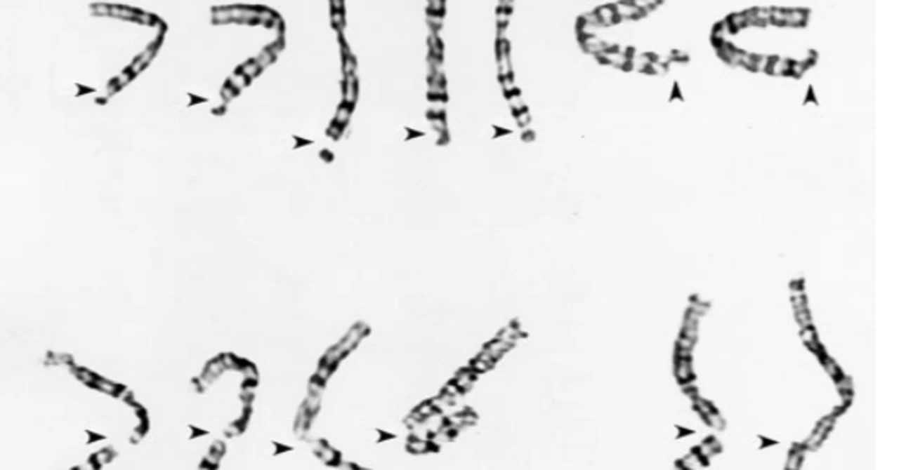
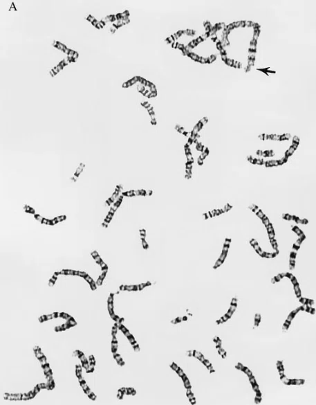
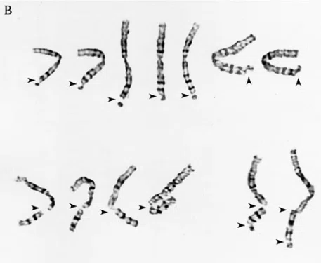
Analysis of primary FFs infected in S-phase with the Towne strain of HCMV revealed a dramatic increase in the incidence of specific breakage on the long arm of chromosome 1. Approximately 16% of the infected cells showed a specific break at either position 1q42 or 1q21, and this was virtually never seen in the mock-infected samples. Fig. 1 shows examples of the typical damage observed in HCMV-infected cells at metaphase (including breaksy gaps in band 1q42, breaks in 1q21, and 2 examples of double 1q21y1q42 breaks).HCMVのTowne株をS期に感染させた一次FFを分析したところ、1番染色体の長腕における特異的切断の発生率が劇的に増加した。 感染細胞の約16%が1q42または1q21のいずれかの位置で特異的切断を示した。 図1は、HCMV感染細胞のメタフェースで観察された典型的な損傷の例である(1q42の切断、1q21の切断、1q21y1q42の二重切断の2例を含む)。
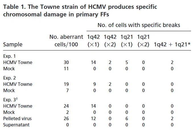
Exps. 1 and 2 of Table 1 summarize the data obtained with the Towne strain of HCMV. Several important features (that also hold true for the results presented in the subsequent tables) should be pointed out. In both mock- and HCMV-infected samples, cells were scored as aberrant if they possessed one or more breaks, gaps, or deletions. At the 1q42 position, the vast majority of damage was in the form of single chromatid breaks, while damage at the 1q21 position was almost equally divided between chromatid and chromosome breaks. The 1q42 break was observed in all viral samples. However, the 1q21 break, although reproducible, was not detected in every trial. Although the preponderance of cells incurred a break in only one copy of chromosome 1, breakage of both copies was present at a low frequency. The dual combination of 1q21 and 1q42 breaks within the same cell (and even the same chromosome) was also sporadically observed. Although aberrations were observed in the mock samples, the damage never included the specific 1q42 break and only once included the 1q21 break (Exp. 9).表1のExps. 表1の1および2は、HCMVのTowne株を用いて得られたデータを要約したものである。 いくつかの重要な特徴が指摘される。 模擬サンプルとHCMV感染サンプルの両方で、細胞は1つ以上の切断、ギャップ、欠失を持つ場合に異常と判定された。 1q42の位置では、損傷の大部分は単一染色分体切断の形であったが、1q21の位置での損傷は染色分体切断と染色体切断にほぼ等しく分けられた。 1q42の切断は全てのウイルスサンプルで観察された。 しかし、1q21の切断は再現性はあるものの、すべての試験で検出されたわけではない。 大部分の細胞で1番染色体の片方のコピーにのみ切断が生じたが、低い頻度で両方のコピーの切断が認められた。 同一細胞内(さらには同一染色体内)で1q21と1q42の二重の切断も散発的に観察された。 模擬サンプルでは異常が観察されたが、損傷が特定の1q42切断を含むことはなく、1q21切断を含むことは1度だけであった(Exp.9)。
-精製ウイルス粒子が1番染色体切断を誘発
When conducting studies on virusyhost cell interactions, it is important to determine whether the virus itself or some component of the infected cell supernatant is responsible for the observed result. To answer this question, we purified virions from the cell supernatant by using a series of high-speed centrifugation and washing steps (see Materials and Methods). Exp. 3 in Table 1 documents that purified virions, and not the infected cell supernatant alone, were capable of inducing the specific chromosome 1 breaks.ウイルスと宿主細胞の相互作用に関する研究を行う場合、観察された結果の原因がウイルスそのものにあるのか、それとも感染細胞上清中の何らかの成分にあるのかを明らかにすることが重要である。 この疑問に答えるため、我々は一連の高速遠心分離と洗浄工程を用いて、細胞上清からビリオンを精製した(材料と方法を参照)。 表1のExp.3は、感染細胞上清だけでなく、精製ビリオンが特異的な1番染色体切断を誘導できることを示している。
-1番染色体の損傷は細胞種特異的ではない
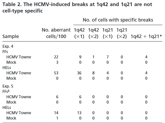
Because primary cells must be used for HCMV infections, there is a risk that underlying fragility in a single individual’s chromosomes will be revealed on exposure to a potentially damaging agent. We therefore tested a completely unrelated source of human embryonic lung cells, also of primary origin. The data in Table 2 clearly demonstrates that the 1q42 break was observed in both trials at comparable or higher levels than in FFs. In Exp. 4, there was also a high incidence of 1q21 breaks over background. These results eliminate the possibility of cell-type specificity.HCMV感染には初代細胞を使用しなければならないため、潜在的にダメージを与える薬剤に暴露されると、一個人の染色体に潜む脆弱性が明らかになる危険性がある。 そこで、全く無関係のヒト胚性肺細胞(これも一次由来)を用いた試験を行った。 表2のデータから明らかなように、1q42の切断は両試験ともFFと同等かそれ以上のレベルで観察された。 実験4では、1q21切断もバックグラウンドより高い頻度で観察された。 これらの結果から、細胞型特異性の可能性は排除された。
-1番染色体損傷は系統特異的ではない
HCMV strains vary considerably in the length of time that they have been continuously cultured in the laboratory and to a lesser extent in their genotype. To assess the role of viral passage number and strain type in the occurrence of the induced 1q42 and 1q21 breaks, we compared the high passage Towne and AD169 strains to the Toledo strain of HCMV. Although slightly adapted to tissue culture, the Toledo strain is genotypically and phenotypically much closer to standard clinical isolates, as its genome possesses a 13-kbp region absent from the high passage laboratory strains (33). As shown in Table 3, both the AD169 and Toledo strains produced results identical to the Towne strain with respect to the incidence of the specific chromosomal breakage in region 1q42, thus discounting any tissue culturey strain-specific phenomena in the induction of this break. The AD169 strain also induced the break at 1q21 in both trials performed.HCMV株は、実験室内で継続培養された期間や、遺伝子型によってかなりの違いがある。 誘発された1q42および1q21切断の発生におけるウイルス継代数と株タイプの役割を評価するために、高継代Towne株とAD169株をHCMVのToledo株と比較した。 Toledo株は組織培養に若干適応しているが、ゲノムの13kbp領域が高継代実験室株にはないため、遺伝子型的にも表現型的にも標準的な臨床分離株にはるかに近い(33)。 表3に示すように、AD169株とToledo株はともに、1q42領域における特異的染色体切断の発生率に関してTowne株と同じ結果を示したので、この切断の誘発における組織培養株特有の現象は否定された。 AD169株は両方の試験で1q21に切断を誘発した。
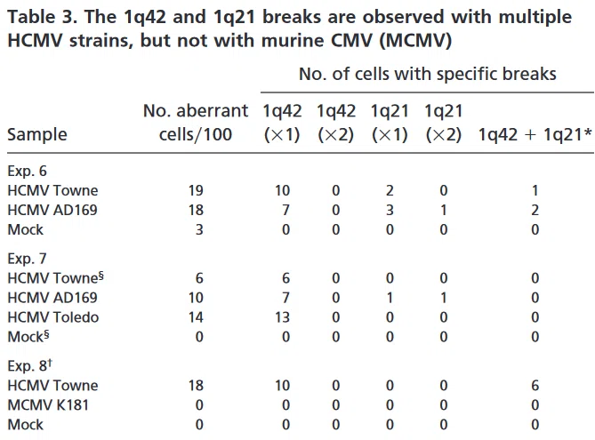
In a related experiment, we tested murine cytomegalovirus (MCMV), which has the ability to infect human cells and initiate viral protein synthesis (34), to see whether induction of damage was a virus-specific phenomenon. As shown in Table 3, Exp. 8, MCMV was incapable of causing chromosomal damage in human cells.関連する実験として、ヒト細胞に感染してウイルス性タンパク質合成を開始する能力を持つマウスサイトメガロウイルス(MCMV)を試験し(34)、損傷の誘導がウイルス特異的現象であるかどうかを調べた。 表3のExp.8に示すように、MCMVはヒト細胞で染色体損傷を起こすことができなかった
Viral Protein Expression Is Not Required for Induction of Damage.
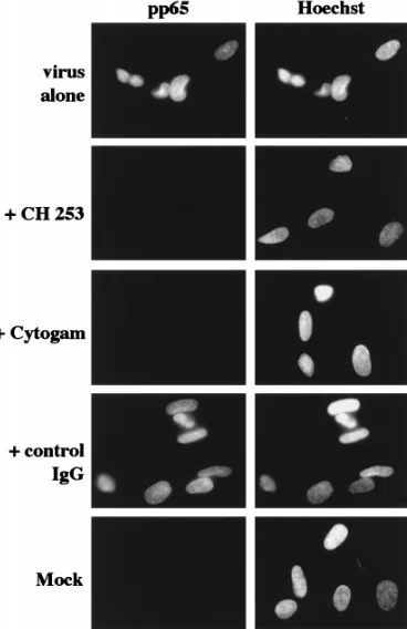
Next, we assessed the requirement for de novo viral gene expression in the induction of the specific breaks. Two parallel virus aliquots (equivalent to an moi of 5) were thawed; one was UV-inactivated, while the second was left untreated. Both UV irradiation procedures and doses (described in Materials and Methods) were predetermined to decrease expression of the HCMV genome, such that ,1% of the cells synthesized viral immediate early proteins at 24 hpi (as assayed by immunofluorescence and demonstrated in Fig. 2).
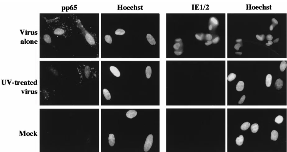
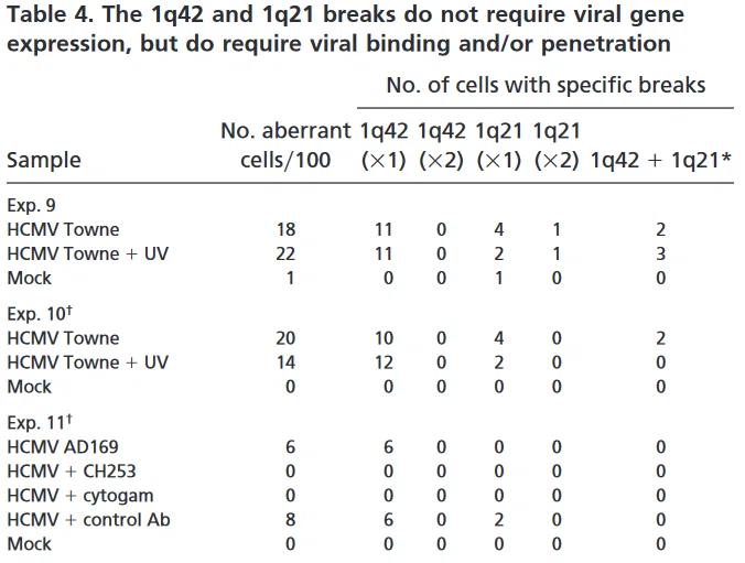
Under these conditions, viral entry was not affected, as measured by pp65 tegument protein staining in the nuclei of all cells, as was observed for cells incubated with untreated virus in this and all other experiments. The addition of sodium pyruvate to the media after irradiation was essential for survival of mitotic cells, as cultures with media lacking this supplement showed apoptotic destruction of all M-phase cells, most probably because of oxygen free radical production during irradiation. Mock supernatant was also treated to control for variability introduced by the irradiation procedure. As can be seen in Table 4, Exps. 9 and 10, both untreated virus and virus UV-inactivated with either irradiation procedure scored equally well with respect to induction of the specific 1q42 and 1q21 damage. Thus, new viral gene expression is not required for the induction of chromosome 1 breaks.
Virion Binding and/or Entry Is Required for Damage Induction.
To determine the nature of the virusyhost cell interaction required for the induction of chromosome 1 breaks, we utilized the following neutralizing Abs: a mAb (CH 253) specific for the viral envelope glycoprotein gB, which is a main component in viral attachment and penetration, and a purified IgG fraction of polyvalent hyperimmune sera isolated from HCMV-positive individuals (Cytogam). CH253 does not block viral binding to the cell surface, but does inhibit penetration of the virus into the host cell (35). As described above, viral penetration was assessed in this experiment by immunostaining with an Ab specific for the pp65 tegument protein. Fig. 3 shows that viral entry is blocked by incubation with the neutralizing Abs, but not with control IgG. As can be seen in Table 4, Exp. 11, both test Abs were equally potent at preventing the induction of chromosomal damage by strain AD169 (and the Towne strain; data not shown). These data suggest that virion penetration, andyor a specific receptoryvirion protein interaction at the cell surface (blocked by the neutralizing Ab), are required for chromosome 1 breakage to occur in S-phase-infected fibroblasts.
考察
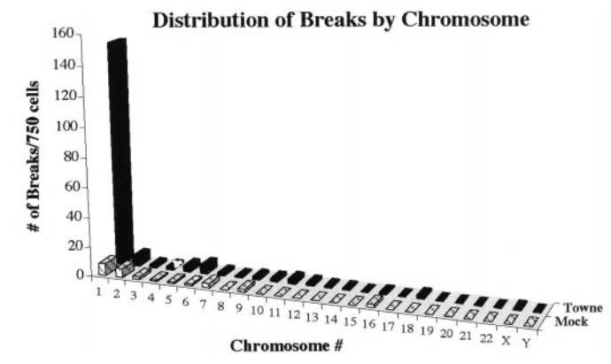
One might expect, in light of the wealth of data concerning other viruses, to see an increased incidence of breakage in all chromosomes in HCMV-infected cells. However, if one compiles the data from all experiments using untreated Towne virus on FFs and examines the distribution of breaks within the aberrant cells, a striking pattern is revealed. As illustrated in Fig. 4, there appears to be a nonspecific, random distribution of low background breaksygaps occurring in the virally infected cells that is parallel to that seen in the mock cells. However, HCMV causes a dramatic increase in the number of breaks in chromosome 1, almost completely accounted for by specific breaks at 1q42 and 1q21. The data show that there is not an increased incidence in overall breaks spread throughout the genome in the HCMVinfected cells, but rather an enhanced fragility at specific loci on chromosome 1.
Why is this the first time that experiments using HCMV-infected cells have revealed specific damage to chromosome 1? First, our studies were performed at mois sufficient to ensure that all cells were infected simultaneously (as seen in Fig. 2). It may be that a higher concentration of viral particles is needed to observe specific damage. In fact, others have noted that not even nonspecific damage was significantly increased above background until mois greater than 1 were used (3). Secondly, and perhaps most importantly, the infections were carried out when a large proportion of cells (approximately 50–60%) were in the S- and G2yM-phases of the cell cycle. At these times, the chromosomes are likely most susceptible to damaging agents resulting from the partial unwinding that occurs during S-phase and the partial condensation events that occur during the G2yM-phases. The preponderance of chromatid vs. chromosome breaks at the 1q42 position would also argue that specific chromosome 1 damage is incurred after DNA replication rather than during G1 (36). In addition, because our experimental protocol resulted in a much larger proportion of mitotic cells at the time of observation, the probability of being able to detect specific chromosomal breaks was enhanced. The cells were harvested fairly rapidly after infection to maximize the potential for seeing damage incurred in the first round of division. Previous analyses were performed much later in infection, when there was likely more global damage to the host DNA, as well as loss of a significant fraction of cells because of virally mediated cell death.
A major question for the future is: what genes lie at these breakpoints? The possible targets residing near 1q42 include: the ADPRT locus involved in DNA repair and replication (37); a potential tumor suppressor gene, whose deletion has been connected to the development of gliomas (38); the major 5S rRNA locus (10); and the USH2A gene (39, 40). The possible targets that reside near 1q21 include: a different proposed tumor suppressor gene deleted in several primary breast tumors (41), the PSU1 small nuclear RNA locus (7), and the DFNA7 gene (42). Two of these loci, DFNA7 and USH2A, are of particular interest with respect to HCMV pathogenesis. The DFNA7 gene located within 1q21–23 has been linked to the inheritance of an autosomal dominant, nonsyndromic, progressive hearing loss (42). The deletion of the DFNA7 gene because of HCMV-induced breakage could potentially be linked to the development of the progressive hearing loss observed in infants congenitally infected with HCMV. The USH2A gene, located at 1q41, close to the most prevalent HCMV-induced break, encodes a protein involved in the development of Usher’s Syndrome Type II. Usher’s Syndrome is an autosomal recessive disorder that affects 3–6% of children born with hearing impairments and is the most frequent cause of combined deafness and blindness in adults (39, 40). These individuals have sensorineural hearing deficiencies at birth and later develop retinal problems. This disease profile is similar to that of children congenitally infected with HCMV. The predicted ORF for the USH2A protein contains both laminin epidermal growth factor and fibronectin type III motifs, and thus the gene may encode a basement membranetype protein. One could envision a scenario where HCMV-induced breaks at this locus caused deletion of the 1q terminal region (in one or both copies of chromosome 1), which was then passed on to daughter cells. Subsequent deletion of the other USH2A locus, either by loss of heterozygosity or chromosome erosion could abolish all USH2A protein expression, producing an Usher-type syndrome similar to congenital HCMV infection.
In future studies, we will examine cells of neuronal and glial descent. The fetal brain is most susceptible to HCMV-induced damage during the first half of pregnancy, a time when neuronal cell division and differentiation are maximal (12). We know from tissue culture experiments that several cell types within the brain show varying degrees of permissiveness for the HCMV infection, and this susceptibility appears to depend on the origin of the cell and the state of differentiation (43–46). The gamut of permissiveness ranges from the complete susceptibility of human primary retinal glial cells to a total block in viral protein synthesis and DNA replication in undifferentiated glioblastoma cells (43, 45). It is interesting to note that the same cell line can become more fully permissive on differentiation (44, 46).
The observation that chromosomal damage can occur in the absence of de novo viral gene expression becomes very important when taken in the context of the variability of permissiveness and restriction to gene expression observed in different neuronal cell types at various stages of differentiation. These data suggest that in utero, semi- to nonpermissive undifferentiated neural cells might be infected by HCMV and incur chromosomal damage. As these neuronal precursors divide, they could then pass on this damage to daughter cells, which in turn could either divide again or differentiate and subsequently migrate to their positions within the cortex or other regions of the developing brain.
Lack of a requirement for viral protein synthesis also sets HCMV apart from other damage-inducing viruses. Prior studies have shown that the adenovirus type 12 E1B protein is absolutely required for induction of damage in infected cells, and can actually induce damage independently of viral infection (9, 47). In addition, the damage induced by the HCMV-related herpes simplex virus, which appears as the uncoiling of chromosome 1q12–21 and the pericentric regions of chromosomes 9 and 16 rather than specific breakage, also requires immediate early viral protein synthesis (48, 49).
What, then, is the mechanism behind this HCMV-induced chromosome damage? One possibility could be the early physiological changes brought about by viral binding and the rapid yet transient induction of c-fos, c-jun and c-myc mRNAs, which occurs even in the absence of serum and with UV-inactivated virus (19, 23). The underlying mechanism also could be related to the early up-regulation of another class of mRNAs that are a subset of genes normally induced by a interferon in uninfected cells (50, 51). This HCMV-associated induction appears to simply require the exposure of the cell to virions, noninfectious enveloped particles, or dense bodies. Viral binding also triggers the generation of reactive oxygen intermediates in some cells, which could potentially lead to DNA damage (52, 53). Alternatively, a viral or cellular protein component of the incoming virion or the viral DNA itself may be responsible for the induced damage. Whatever the mechanism, it is clear from our experiments that HCMV infection, without the requirement for new viral gene expression, sets the stage for specific damage to chromosome 1. Thus, any or all of these early events could lead to the induction of a DNA damage pathway or to a block in DNA repair processes in which chromosome 1 is particularly susceptible. The challenges are now to decipher the signaling pathway leading to chromosome damage and to assess the importance of these breaks in chromosome 1 for the pathogenesis of HCMV in the human embryo during development.
This work was supported in part by National Institutes of Health Training Grant AI 07036, March of Dimes Grant 5-FY98-0727 (to E.A.F.), and National Institutes of Health Grant CA73490 (to D.H.S.).
いいなと思ったら応援しよう!
 全記事無料公開しつつサポートのみでの生存を目指す男です。
何か響くものがあれば、期待できる何かがあれば投げ銭頂けると幸いです。
よろしくお願いいたします。
全記事無料公開しつつサポートのみでの生存を目指す男です。
何か響くものがあれば、期待できる何かがあれば投げ銭頂けると幸いです。
よろしくお願いいたします。